- Home
- Knowledge library
- Non-infectious claw horn lesions in cows
Non-infectious claw horn lesions in cows
These lesions are caused by imbalances of weight, trauma to the foot and other physical factors.
Back to: Lesions of cows’ feet
Success factors
Non-infectious claw horn lesions can usually be treated and improved by using success factors 2, 3 and 4.
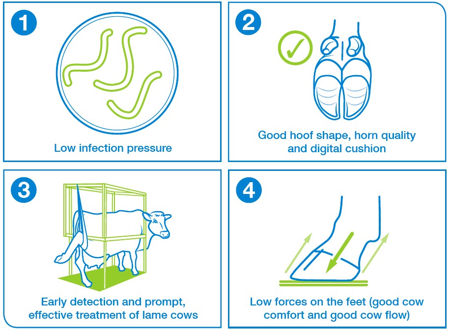
Non-infectious lesions can also be complicated when infections are introduced. Our guides for identifying mixed infections and advice on when to consult a vet can help.
Treating non-infectious claw lesions will usually rely on success factors 2, 3 and 4, so refer to our guides on treating non-infectious lesions like sole bruising and blocking a foot while treating lameness.
Sole haemorrhage (SH)
Description
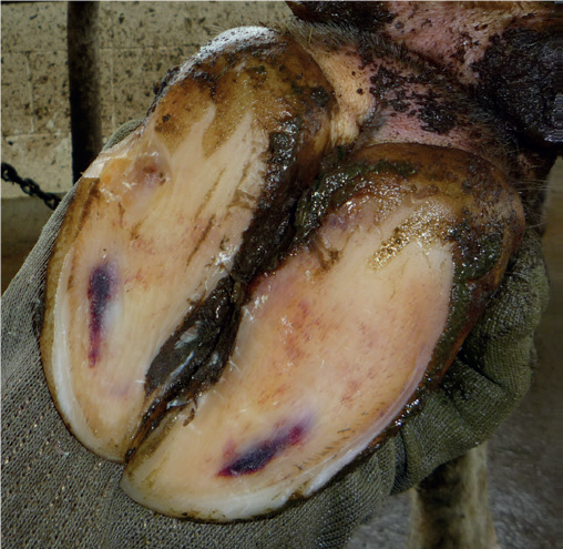
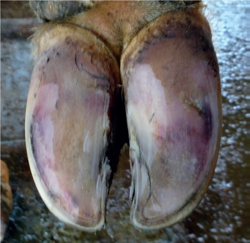
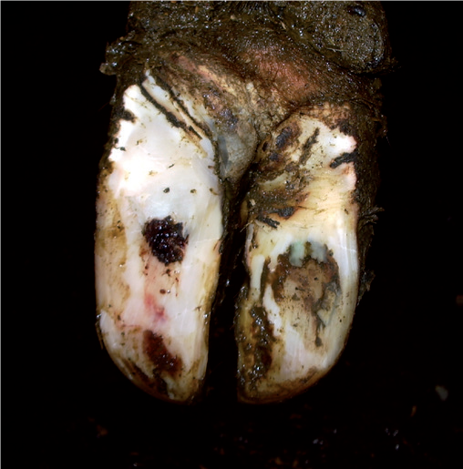
Sole haemorrhage, also known as sole bruising, is characterised by the presence of red and sometimes yellow marks or areas on the sole, and may occur where the sole is particularly thin.
Sole haemorrhage is due to pressure on the corium, leading to damage (inflammation) to cells and blood vessels. The corium is the tissue which produces new sole horn, and is sandwiched beneath the pedal bone and the sole horn itself.
Whilst the bruising or discolouration seen on the sole was sometimes described as “laminitis”, this is not really the best term to use.
There are many risk factors for excess pressure leading to cellular damage and sole haemorrhage. A common one is excess standing time, for example if cow comfort is sub-optimal, if sheds are crowded or if cows spend a long time waiting to be milked.
Other factors include cows walking long distances, or walking surfaces being particularly uneven (e.g. very bumpy or broken concrete).
An additional risk is that, for a few weeks around calving, the pedal bone sinks slightly within the hoof. This further increases pressure on the corium. Providing extra care for fresh-calved cows so they lie down for longer (for example, loose housing and short milking-times) can help protect these at-risk cows and heifers.
More recent research shows the importance of the digital cushion, which is a pad of fat sandwiched under the pedal bone, and which normally acts to dissipate the forces whilst cows stand on hard surfaces, or walk long distances. In many ways, the digital cushion can be considered like the air-pockets in air-cushioned soles of some footwear. Thin cows have thinner digital cushions, and they seem less dense too. This means thin cows have less protection from concussive forces, and they are more likely to suffer from sole haemorrhage. This is probably the biggest influence that nutrition has on lameness: anything that results in cows losing weight or being thin is a big risk for this type of foot damage.
Put all of these risks together, and it helps explain why fresh calved heifers, losing weight and not lying down in cubicles for as long as they should are particularly prone to this condition on some farms.
A vicious cycle can follow sole haemorrhage, because inflammatory chemicals can cause permanent changes to the pedal bone, and possibly the structure of the digital cushion, leading to further episodes of bruising. Sole haemorrhage is the usual precursor to sole ulcers.
- In very mild forms, the sole discoloration is yellow to pink
- More severe is red to purple
- Caused by damage to the corium (pressure), leading to leaking serum or blood being incorporated into new sole horn
- Discolouration towards the toe, or even the entire sole, often points to the sole being too thin
Typical risks
- Poor acclimatisation to concrete floors and/or cubicles
Associated success factors: 2 and 4 - Too much time standing and poor cow flow – see WLD and SU
Associated success factors: 4 - Thin soles
Associated success factors: 2 and 4 - Possibly dietary factors; loss of support for pedal bone; reduced digital cushion (thin cows/weight loss); possibly acidosis leading to biotin deficiency
Associated success factors: 2
Sole ulcer (SU)
Description
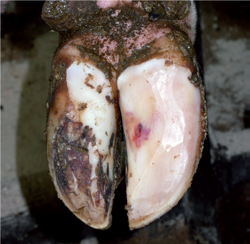
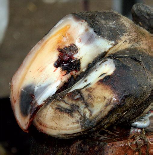
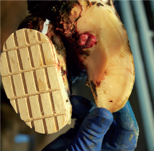
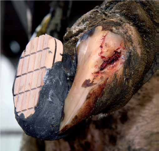
Sole ulcers are a very painful type of non-infectious hoof lesion - usually located where the sole and heel bulb meet. They arise when the soft tissues inside the sole are damaged and normal horn cannot be produced, exposing sensitive tissues to the environment and can cause severe mobility problems in dairy cattle. Sole ulcers are a more severe manifestation of sole haemorrhage. The risk factors are therefore similar.
There is usually a time lag between the initial trauma and the development or observation of the sole ulcer lesion. Sole ulcers are often seen directly under a particular pressure point towards the rear of the pedal bone. Sole horn grows at a rate of around 5mm per month; but hoof horn grows at a rate of around 5mm per month; the sole of the hoof is approximately 10-15mm thick, thus the trauma event causing the ulcer is likely to have occurred two to three months earlier.
Identified and treated sooner, the condition could usually have been “nipped in the bud” as a less severe sole haemorrhage lesion. Treating by taking weight off the affected claw and the use of anti-inflammatory drugs would have prevented the condition progressing to an ulcer.
Sole ulcers commonly affect one or both of the outer hindclaws, particularly in bigger, high-yielding dairy cattle kept under conditions where cow comfort is poor. Where both hind feet are affected it isn’t always easy to tell which hind foot the cow is most lame on, and cases may be missed.
If the corium is exposed, infection can enter the deeper structures of the claw and spread, causing serious problems and allowing deep infections to develop. Cows which have progressed to having a sole ulcer lesion will almost always have sustained irreversible damage to the inner structures of the hoof, including the pedal bone, due to inflammatory chemicals. Therefore, even if the initial treatment is successful, cows which have suffered a sole ulcer once will usually suffer from the condition repeatedly throughout the rest of her life.
- A pressure point exists towards the back of the sole, leading to poor horn formation and bleeding in the horn. Sole ucers progress from sole haemorrhage
- The ulcer develops from within the deeper layers of the sole. Once the outer sole horn has been removed, flesh (the corium) can be seen protruding through the ulcer site
- When present, sole ulcers are often on outer claws of both hind feet
Typical risks
- Excess time standing; poor cubicle comfort; long milking times; long lock-up times; overcrowding; heat stress
Associated success factors: 4 - Thin cows; cows losing weight after calving; old cows with less shock-absorbing capacity from the digital cushions
Associated success factors: 2 - Poor support of pedal bone, for example around calving period; possibly dietary factors too
Associated success factors: 2 - Overgrowth of sole thickness; excessive wall abrasion/abnormal wear, often associated with concrete floors
Associated success factors: 2 - Long toes; eroded heels; poor foot/leg conformation
Associated success factors: 2 - Not enough bedding; poor grip on cubicle surface; too-small cubicle dimensions for size of cow and lack of cubicle training
Associated success factors: 4 - Poor attention to fresh-calved cows and heifers, for example, hierarchical stress
Associated success factors: 4 - Slow detecting and treating early lameness (at bruising stage)
Associated success factors: 3 - Incorrect foot-trimming method
Associated success factors: 3 - Previous inflammation in the foot causing bony changes, and possibly hardening of the digital cushion; for example, delayed treatment or failure to use NSAIDs in treatment of early sole ulcers
Associated success factors: 3
White line disease (WLD)
Description
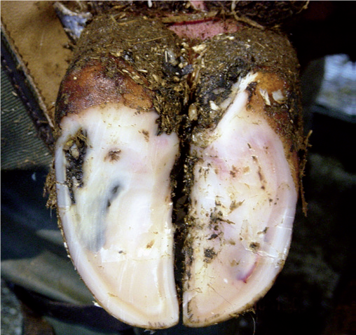
White line disease is a non-infectious condition that occurs when the sole separates from the side wall of the hoof, allowing foreign material to penetrate and infect the white line region.
The white line is easily damaged and is often an entry point for infection; tracks of infection may cause a localised abscess, or may penetrate to form a deeper abscess, and solid foreign bodies may lodge in the softened, widened area and may push through to the sensitive corium beneath. Exposure to moisture softens the region still further and any rupture of the structure is worsened by the impact of movement, particularly among animals housed on concrete.
- White line disease is a major cause of lameness with the incidence in older cows being as high as 35%
- The outer claw of the hindfoot is usually involved; where both claws are affected the problem may escape notice until lameness is more obvious in one limb than the other
- The degree of pain and lameness depends on the rate of development and the extent of the abscess; a discharge of pus from where the skin and horn meet is always reason to suspect a white line lesion and the white line should always be examined very carefully in cases of lameness with these symptoms
- The region of the wall of the hindlimb most often affected is the area of the claw that absorbs the concussion from walking first and in which horn growth and wear occur the most
- Exposure to moisture from wet conditions underfoot softens the tissues still further.
- In mild cases, the horn of the wall can be seen separating slightly from the horn of the sole, at this junction, known as the white line. Sometimes, there is blood staining (bruising)
- Pus can track up the wall and burst at the coronary band or under the sole to burst at the heel (creating a double sole)
Typical risks
- Poor grip on floors; sharp turns; overcrowding and dead-end passages; bulling cows, no loafing area
Associated success factors: 4 - Stockmanship factors; rushing along tracts, pressure during herding and rough use of backing gate; poor cow flow in collection yard
Associated success factors: 4 - Poor tracks: wet, stony ground; long distances and long standing times
Associated success factors: 4 - Rough concrete; new concrete (due to being rough, but also chemical horn damage)
Associated success factors: 4 and 2 - Poor acclimatisation to concrete floors
Associated success factors: 2 - Weak horn: e.g. nutritional imbalance (for example, biotin deficiency); wet feet
Associated success factors: 2 - Thin soles: e.g. over-trimming; too much abrasion; walking long distances
Associated success factors: 2
 resaved.jpg)
Useful links
Lameness in cows: when to involve the vet
Lesion recognition and trouble shooter guide
If you would like to order a hard copy of the Lesion recognition and trouble shooter guide, please contact publications@ahdb.org.uk or call 0247 799 0069.

